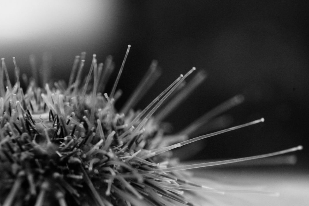
“The white sea urchin (Lytechinus pictus) is found below the tide line,” writes marine biology graduate student Julia Notar in her submission. “I study how these animals see, and what they can see. They usually live in flat, sandy areas, where there aren't many places to hide from fish predators. Different species of sea urchins, which live in rocky areas, usually hide from fish in dark crevices in, between, or under rocks. Those urchins can use their blurry, but still useful vision to find those hiding spots. Does this species, which doesn't live in an environment with many hiding spots, do the same thing?”
Julia Notar, courtesy “The Art of a Scientist”
/
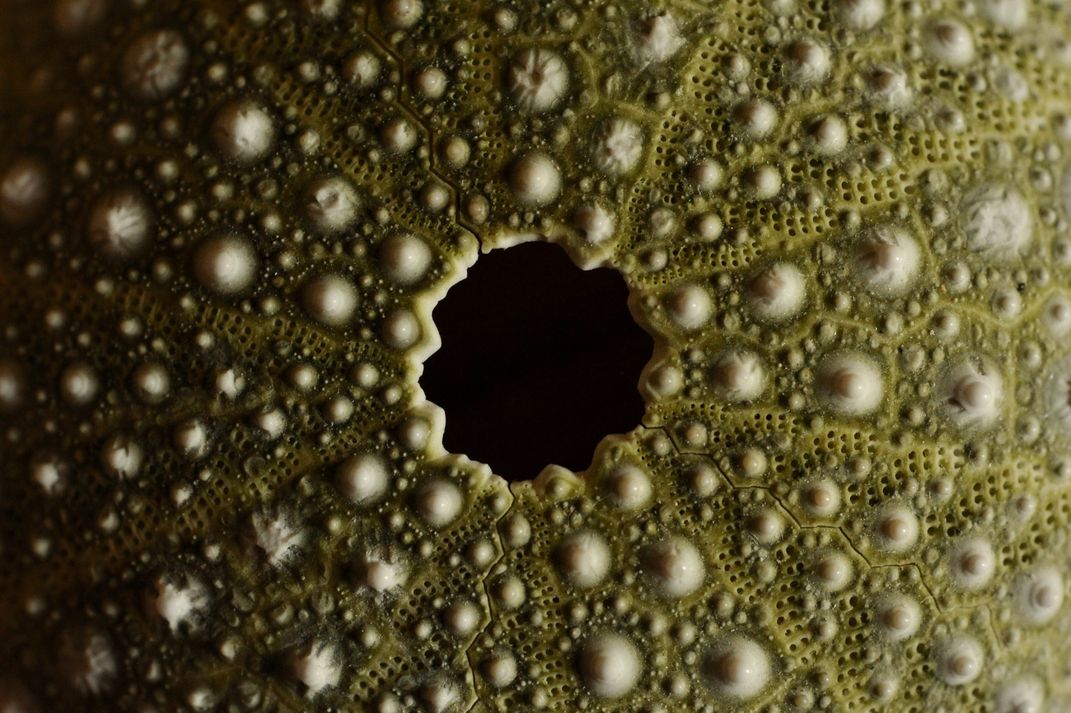
The internal skeleton of a purple sea urchin (Strongylocentrotus purpuratus). “These animals don't have eyes, but we think they are using skin all over their skeleton, separated by their spines, to sense light and shadows. Looking at the skeletons of different species lets us calculate the size of the pixels and estimate how good their vision might be,” writes Julia Notar.
Julia Notar, courtesy “The Art of a Scientist”
/
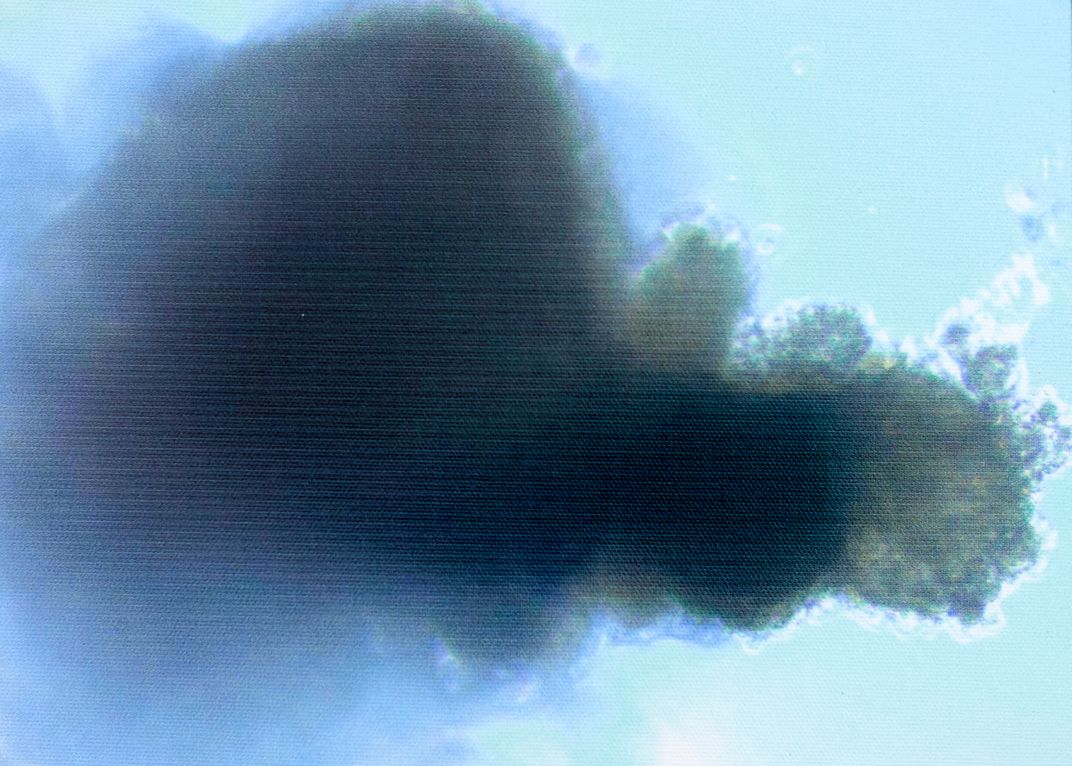
“Cancer is a dreaded diagnosis, and no cancer patient is happy to see what their intruder looks like,” writes pathology PhD student Larisa Gearhart. “But to a scientist, a tumor cell cluster like the one shown here, made from an aggressive human breast cancer grown in a lab, is a fascinating enigma. These clusters and their inner workings hold the secrets to how the tumor will grow, evolve and spread.”
Larisa Gearhart, courtesy “The Art of a Scientist”
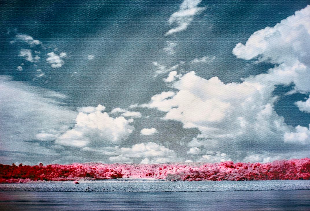
“This photograph was shot in southeastern Peru, near the headwaters of the Amazon,” writes geology graduate student Wout Salenbien. “Infrared light, invisible to the human eye at frequencies ranging from 700 to 900 nanometers, is strongly reflected by the chlorophyll inside plants cells whereas most other materials have a much more muted response. Using an infrared filter on your camera, it is possible to capture images in this invisible landscape of light. Using false color processing techniques on the highly reflective foliage, it is possible to separate a range of bandwidths that correspond to varying chlorophyll concentrations and assign a different color to those values. As such, the more intense the pink color is in the picture, the higher the concentration of chlorophyll. You’ll notice that not every tree has the same amount of pink, which gives you an indication of the plant’s health.”
Wout Salenbien, courtesy “The Art of a Scientist”
/
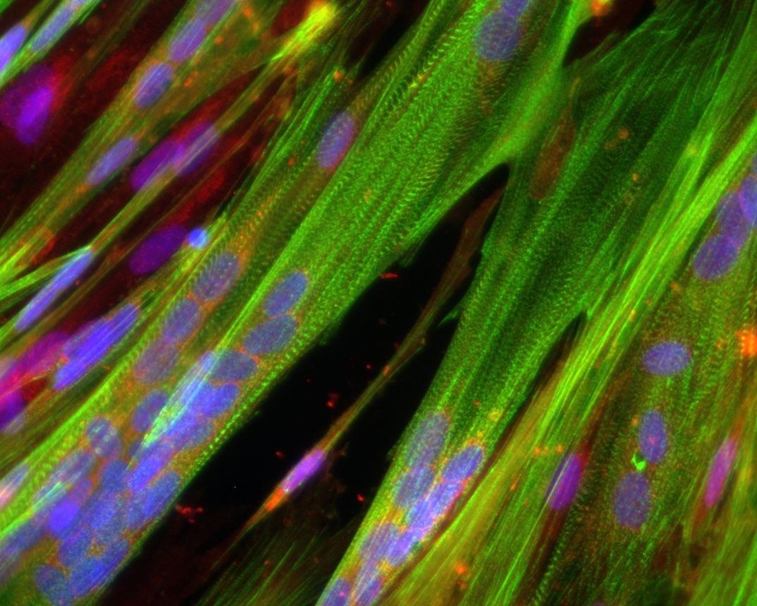
“This photo shows striated human skeletal muscle myotubes – the building blocks of strength and movement in the human body,” writes biomedical engineering student Megan Kondash. “Each cluster of blue nuclei represents a group of formerly individual cells which have fused together to create a unit capable of contraction.”
Megan Kondash, courtesy “The Art of a Scientist”
/
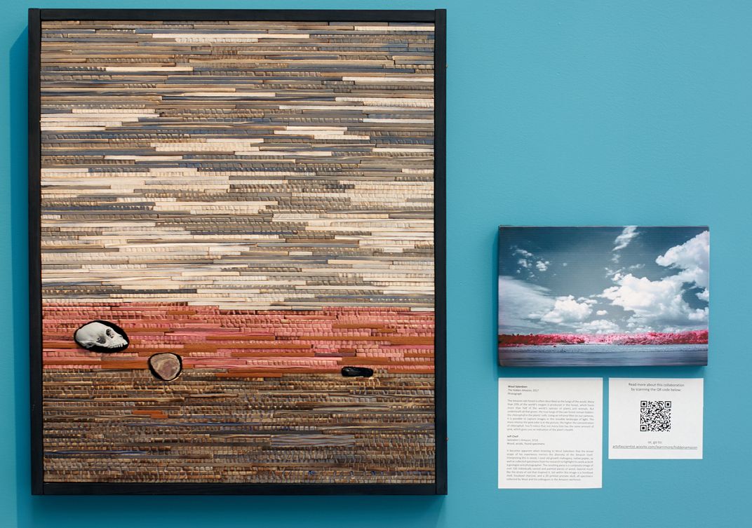
“It becomes apparent when listening to Wout Salenbien that the broad scope of his experience mirrors the diversity of the Amazon itself,” writes artist Jeff Chelf. “I used old growth mahogany, native polar as well as collected specimens from his research to highlight his work as both a geologist and a photographer.”
Jeff Chelf, courtesy “The Art of a Scientist”
/
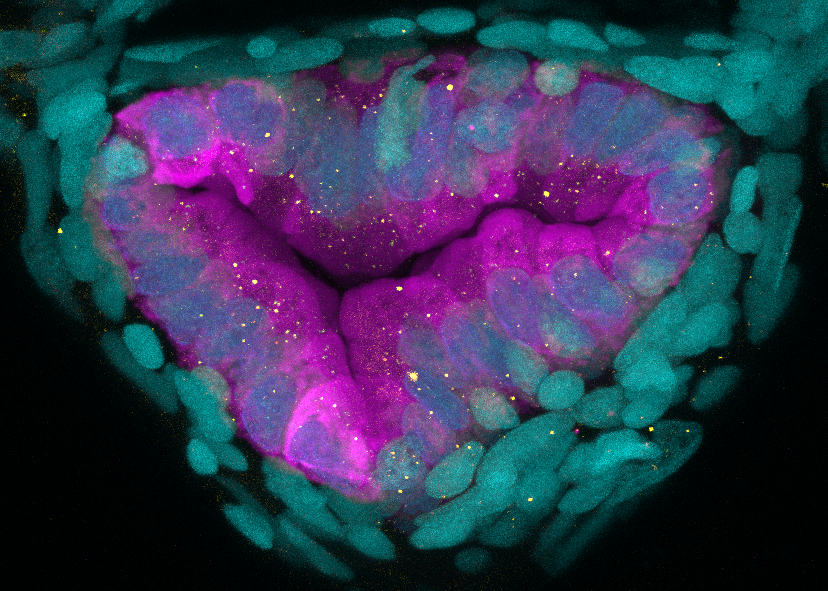
A cross section of the intestine from a zebrafish. “Zebrafish are a powerful model organism, which are using in concert with pharmacological, genetic and gnotobiotic manipulations to gain new insights into how the intestine functions in homeostasis as well as the underlying mechanisms of disease,” writes Ted Espenschied, a graduate student in molecular genetics and microbiology.
Ted Espenschied, courtesy “The Art of a Scientist”
/
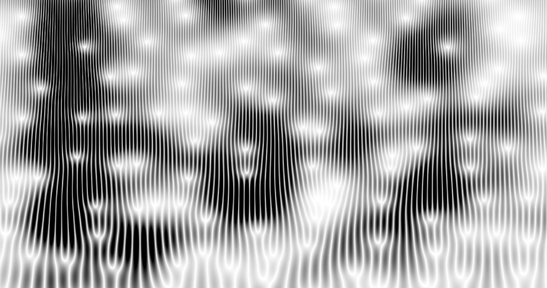
“Time series which describe statistical or natural processes can often have multi-scale periodic structures that are invisible to the human eye.” writes John Malik, a graduate student in mathematics. “A mathematical device called the short-time Fourier transform reveals this structure. The phase information obtained from the Fourier transform is usually ignored, but its contours are visually stunning. This image, entitled ‘Phases of the Heart,’ displays the phase information extracted from an electrocardiogram signal.”
John Malik
/
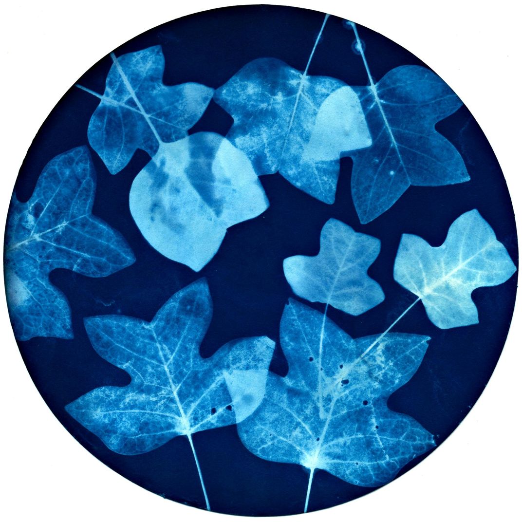
“This cyanotype print captures the essence of the tulip poplar (Liriodendron tulipifera), the tallest native hardwood tree of the Eastern U.S.,” writes artist and research assistant Ansel Oommen. He explains that the cyanotype is an alternative photographic process that relies on the light sensitive properties of two iron compounds. “When exposed to UV light, these compounds form a deep, rich pigment called Prussian blue. Traditionally, items such as leaves were placed on top of paper sensitized with the chemical solutions of these compounds and then exposed to sunlight. Areas that were covered by the items would not form Prussian blue. The resulting negatives were popularized in botany by Anna Atkins, the first female photographer. In 2016, I developed a combination process which chemically pre-treats the leaves to be translucent. As a result, UV light is able to selectively pass through the leaf to varying degrees based on whether it encountered lignin or not. Like cellulose, lignin is a natural polymer that makes wood strong and sturdy. It also helps form the ‘skeleton’ of the leaf. And just like bones on an X-ray, the veins appear more highlighted because they absorb more radiation than the surrounding soft tissue. This print is a testament to the importance of interdisciplinary study. By migrating between botany, alternative photography, radiology, and photochemistry, I was able to cross-pollinate them all in the process.”
Ansel Oommen, courtesy “The Art of a Scientist”
/
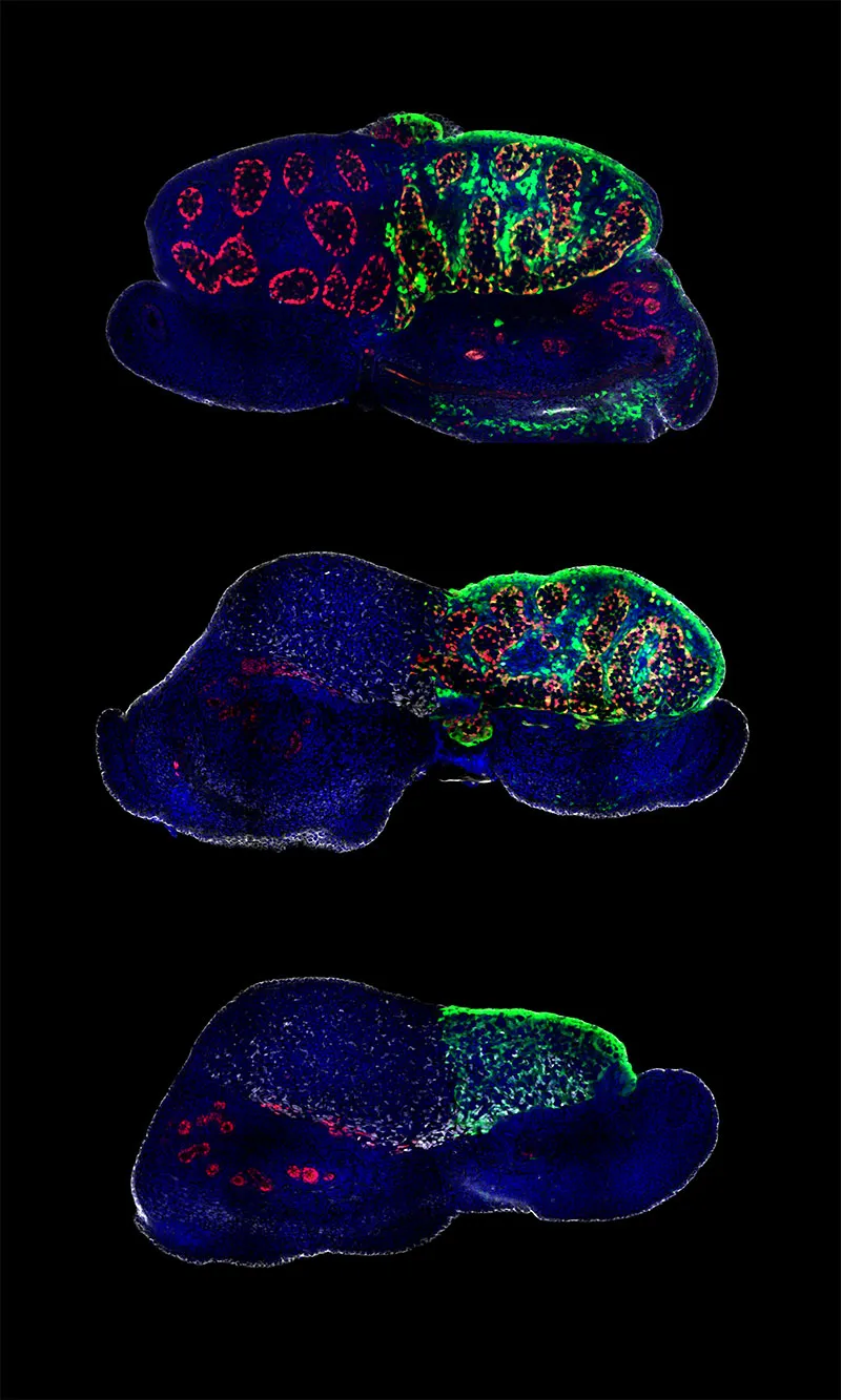
“The mammalian male and female gonad are indistinguishable when they first form,” writes Corey Bunce, who is working toward his PhD in the Developmental and Stem Cell Biology program. “Errors can lead to the development of indeterminate gonads, which contain both testis and ovary parts, and will often later resolve to one type or the other. We seek to understand how the programs underlying the distinct fates influence each other.” He adds, “Here, organs were collected from developing mice soon after the gonad began to take on characteristics specific to a testis or ovary.”
Corey Bunch, courtesy “The Art of a Scientist”
At the Rubenstein Arts Center on Duke University’s campus, an image from a microscope makes an alien landscape out of the knobby, radial symmetry of a sea urchin skeleton. Turquoise ovals crowd a ring of fluorescent magenta flesh in another image—a section of intestines inside a zebrafish. And monochromatic points of light float in front of a set of black and white lines in what could be an abstract work of art. The image is actually the electrical signal from a heartbeat subjected to a mathematical process and then made visual.
Thirty-four works created by 22 scientists and 13 artists are now on display in a new exhibition called “The Art of a Scientist” through August 10.
The whole thing arose out of a miscommunication. Duke University PhD student Casey Lindberg was enjoying a downtown art walk in Durham, North Carolina with a friend. She was delighted by the diversity of art around her and mused: “Wow, what if we did an art walk with science pieces?” Her friend thought she meant a collection of artists’ interpretations of science work. But Lindberg was actually dreaming of a display of science images produced in the lab.
Then she realized, why not have both?
Lindberg took the idea to fellow graduate students Ariana Eily and Hannah Devens. The three are co-chairs of the science communication committee for a student group called Duke INSPIRE. The group’s mission is to accelerate academic scientific progress and facilitate public engagement with the scientific process. “We wanted to get scientists and artists working together to kind of show off the different sides of science and art,” says Eily. “To let people see just how connected those two different disciplines are.”
After a year and a half of dreaming, planning and organizing, the trio’s efforts have come to fruition. The group solicited submissions from labs around the university as well as artists’ groups and galleries in the area. Then they paired artists and scientists who wanted to work together. For this first show, they accepted all the pieces submitted.
The three students are no strangers to blending art and science. Lindberg is learning about photography though she spends much of her time researching the long-term effects of pollutants on wild fish populations. Devens’ graphic design skills went into creating the poster for the exhibit. In the lab, she is exploring the genes that shape development and evolution using sea urchin embryos as a model organism. Eily is a self-proclaimed dabbler in “lots of different places.” She sings in a friend’s band, occasionally works as a sous-chef for a catering business and does improv theater. She will defend her thesis this year on the intricacies of a symbiotic relationship between an aquatic fern called Azolla and the cyanobacteria that live within its leaves.
“The thought processes or the way that scientists and artists both approach a question are really similar,” Eily says. “The time that goes into planning out how you get from the conception of an idea to actually getting some sort of physical result and the different trial and error processes that take place to get you there are similar.” She has translated her improv work into coaching scientists on how to hone their speaking skills to communicate about their research.
Some of the pieces in the exhibit are very similar to those that appear in scientific papers— which can hold an unexpected bounty of beauty. “People who are not in the science community might not realize how much of an artistic eye scientists do bring into creating figures,” says Devens. Others arose from artists interpreting scientists’ work. Still others are the result of collaboration.
One photograph by geologist Wout Salenbien captures a South American rainforest, but the foliage is colored different shades of pink and red to highlight the more productive trees. Artist Jeff Chelf then took that color palate and used a variety of South American wood types to create a sculpture image that mimics the look of the rainforest in profile and evokes images of soil profiles. Embedded within the 500 pieces of wood are fossils and a printed replica of a primate skull collected by the geologist and his colleagues while in the Amazon.
At the exhibit’s opening, the artists, scientists and the public all mingled. There, Lindberg noticed that despite stereotypes of both artists and scientists being “odd balls with weird curious habits,” it was hard to tell who was a scientist and who was an artist. “Put everyone in the same room and you can't tell the difference,” she says. “All of our artists and scientists just meld together really well.”
The three plan for the exhibit to become a yearly occurrence. Already they’ve had interest from other artists and scientists who want to be involved in the next installment. They hope that the show sparks interest, especially in children that come to see it.
“There’s the kind of old way of thinking: Are you left-brained or right-brained?” says Eily. “But we just want to show you don’t have to choose one or the other, you can do both.”
“The Art of a Scientist” runs through August 10th at the Rubenstein Arts Center in Durham, North Carolina. Programming is free and includes a Family Day on July 14th with hands-on science activities and a panel discussion on August 4 featuring professionals that blend science and the arts.
Get the latest Travel & Culture stories in your inbox.